
ABOUT IMAGING
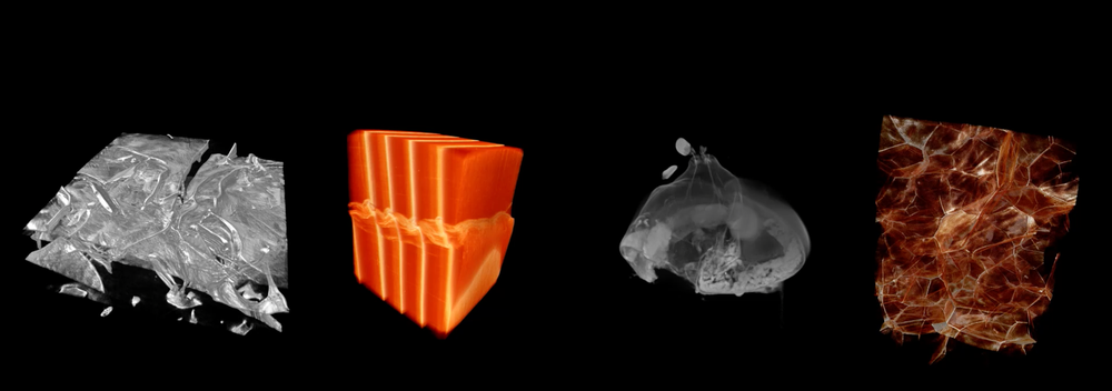
The Imaging theme covered acquisition, processing and applications in imaging that are relevant to systems using synchrotrons and/or neutron sources. The theme considered all possible length scales and subjects accessible with modern and future methods. The focus was on finding new image reconstruction and/or analysis techniques that can help to extract meaningful information from x-ray/neutron imaging data and on connecting different methodologies and competences in order to shed new light on challenges in imaging of different subject matters.
MAJOR EVENTS

CORE GROUP
IPDD WG 3 member and Imaging Core Group Member, LINXS Fellow
Rajmund is a senior scientist, professor at the MAX IV Laboratory (Lund University) and an honorary professor at the Technical University of Denmark. He is a specialist in X-ray physics with emphasis on the enhancement of X-ray imaging for biology and material science applications: development and applications of coherent imaging with improved temporal and spatial resolution.
Imaging Core Group Member, LINXS Fellow
Prof. Jacobsen is the head of the X-ray Science Division at the Argonne National Laboratory and a Professor at the Department of Physics and Astronomy at Northwestern University. With his team, he is focused on developing new methods in x-ray microscopy, and applying them to interesting problems in biology, environmental science, and materials science.
Imaging Core Group Member, External LINXS Fellow
Henning Friis Poulsen is a professor at the Technical University of Denmark (DTU) and heads the 3D imaging centre at there. He is chairman of Danscatt, the Danish instrument centre for neutron and synchrotron users. He is one of two PIs on DanMAX, the Danish beamline at MAX IV, and PI in the societal partnership “Linking Industry with Neutron and X-rays” (LINX - no relation) as well as interim manager of a MAX IV imaging data analysis centre, being co-hosted by DTU and KU.
GUEST RESEARCHERS
Guest researcher at LINXS between Oct 2021 - Nov 2021
Adam Hitchcock was born and educated in Canada (B.Sc., Chemistry, McMaster, 1974; Ph.D., Chemical Physics, UBC, 1978). His research focus is inner shell excitation spectroscopies and spectromicroscopies. A professor at McMaster since 1979, his group has studied inner shell electron energy loss (EELS) spectroscopy of gases and surfaces, using home built instruments. In 1980 he started synchrotron experiments, initially hard X-ray spectroscopy of materials at Cornell (USA), then soft X-ray spectroscopy of gases at LURE (France) and SRC (USA). In 1994 he began developing soft X-ray transmission microscopes (STXM) and photoemission microscopes (PEEM) at ALS (USA). He helped establish the Canadian Light Source (CLS, Saskatoon) and the CLS spectromicroscopy beamline, currently equipped with 2 STXMs and a PEEM. In 2006, he was awarded fellowship of the Royal Society of Canada (FRSC, Canada’s highest scientific honor), for his contributions to development of X-ray microscopy and the CLS. His current research is focused on technique developments of STXM and ptychography and their application to automotive fuel cell materials, in situ electrochemistry, magnetic bacteria, and catalysts for CO2 reduction.
WORKING GROUPS
IMAGING WG 1
Working group ON New Opportunities in Imaging with X-rays and Neutrons
Large Scale facilities such as MAXIV and ESS (as well as other similar facilities around the world) offer great new opportunities for imaging of materials and structures. The aim of this WG is: (i) to inform, educate and develop potential user groups of these possibilities towards establishing a strong user base for the techniques and to bring in new user groups; (ii) to provide a forum for bringing together developers and practitioners of some of these techniques to identify and discuss key areas for development, which could grow into larger activities in their own right. There is a particular focus on methods that are relevant to MAXIV and ESS and a key aim is to enhance the possibilities for new scientific developments and applications at these local facilities, but the discussions should also consider possibilities at other facilities.
A key basis of this WG is the observation that a successful mechanisms to enable new science at large scale facilities has been the tight collaboration between a technique developer and a user group with a challenging case. A case that requires non-standard imaging approaches will trigger creativity in applying and developing new techniques. The WG will help to identify and realize such activities.
X-ray Fluorescence Techniques
Coherent Diffraction
Former LINXS Co-Director and Food WG 3 member, LINXS fellow
Associate professor at the Dept. of Solid Mechanics at the Faculty of Engineering (LTH), where he is also in charge of the 4D-Imaging Lab x-ray tomography facility. Came to Sweden in 2011 after moving from Laboratoire 3R in Grenoble, France.
Imaging Core Group Member, LINXS Fellow
Prof. Jacobsen is the head of the X-ray Science Division at the Argonne National Laboratory and a Professor at the Department of Physics and Astronomy at Northwestern University. With his team, he is focused on developing new methods in x-ray microscopy, and applying them to interesting problems in biology, environmental science, and materials science.
Opportunities in Imaging Working Group member, LINXS Fellow
David Paterson led the development of the Australian Synchrotron’s X-ray Fluorescence Microscopy beamline and has more than two decades of experience in synchrotron radiation research.David has a passionate interest in using X-ray fluorescence microscopy to improve our lives particularly in the fields of environmental science and new materials research.
His research interests are focused on disruptive technology in X-ray fluorescence microscopy, particularly detector and data acquisition schemes. He has fostered and promoted the R&D100 awarded Maia detector at the Australian Synchrotron.
Opportunities in Imaging Working Group member, LINXS Fellow
Karina Thånell is the project manager and scientist for the SoftiMAX beamline at MAX IV Laboratory, Lund University. Her interests lie in Scanning transmission X-ray Microscopy techniques using soft x-rays (up to 2.5 keV), including fluorescence methods, and coherence based imaging, such as ptychography and holography. The beamline is currently being constructed and will be available for users in 2019.
Opportunities in Imaging Working Group member, LINXS Fellow
Beamline scientist at CXI at MAX IV, Lund University.
Opportunities in Imaging Working Group member, LINXS Fellow
Project manager at the NanoMAX beamline at MAX IV, Lund University.
IPDD WG 3 member and Opportunities in Imaging Working Group member, LINXS Fellow
Martin Bech is Senior Lecturer at the division of Medical Radiation Physics, Clinical Sciences, Lund University. He is interested in X-ray phase contrast and dark-field imaging using grating interferometer, Soft tissue biomedical imaging, Micro-CT imaging using synchrotron radiation and x-ray tubes.
Opportunities in Imaging Working Group member, LINXS Fellow
Dr. Edwin Fohtung is an Associate Professor at Rensselaer Polytechnic Institute in New York. He received his Ph.D. ( Dr. rer. nat., summa cum laude) from the University of Freiburg, Germany in 2010. Before he joined Rensselaer Polytechnic as Faculty in 2019 he was the LANSCE (Los Alamos Neutron Science Center) Professor of physics at Los Alamos National Laboratory and New Mexico State University. Before this, he was a postdoctoral fellow with the Department of physics at the University of California San Diego in the Shpyrko Group and worked at the Institute for Photon Science and Synchrotron Radiation in Karlsruhe Institute of Technology (KIT) in Germany. Prof. Fohtung earned his B.Sc. (2005) and M.Sc. (2007) in Technical and Applied Physics from the St. Petersburg State Polytechnic University Russia. Prof. Fohtung is particularly interested in nanoscale structure, fluctuations, and dynamics arising due to coupled and competing spin, orbital, charge, and lattice degree of freedom. To interrogate such order parameters in a variety of condensed matter systems, the Fohtung Research group develops novel synchrotron-based coherent x-ray and neutron scattering techniques. Professor Fohtung is the recipient of numerous awards including the LANSCE Professorship and Rosen Fellow at Los Alamos National Laboratory. He is also the recipient of multiple grants from the US Department of Defense, Airforce of Office of Scientific Research (DoD-AFOSR), the National Nuclear Security Administration (NNSA), the National Science Foundation (NSF) and other LANL directed research and development program awards. Professor Fohtung is currently a visiting professor at Lund University and a LINXS (Lund Institute of Advanced Neutron and X-ray Science) fellow in Lund Sweden.
Opportunities in Imaging Working Group member, LINXS Fellow
Dina Carbone is a beamline scientist of Max IV Laboratory with long-standing expertise in the development of advanced synchrotron radiation methods based on hard (coherent) x-ray nano beams applied to material science. These include, among others, scanning diffraction and inverse microscopy to investigate the correlation between crystal structure and functionality of confined materials.
Dina has joined Max IV in 2014, where she has given a fundamental contribution to the construction of the diffraction endstation of NanoMAX and to the implementation of coherent X-ray based techniques. She recently moved to BLOCH, the ARPES beamline of Max IV, to broaden her scientific expertise.
Dina enjoys supervising young researchers and students that she meets within her international collaborative network, and is interested in scientific outreach. She is co-author of a book on the use of x-ray nanobeams applied to nanoscience.
Imaging Working Group member, LINXS Fellow
Anders Mikkelsen, Director at NanoLund, Lund University, Sweden
IMAGING WG 2
Working group on GeoArCH: Geology, Archaeology and Cultural Heritage
Geological and Archaeological Working Group for the development of imaging using synchrotrons and neutron sources. The new large infrastructures at LU (MAXIV and ESS) will play an important role to merge interests across faculties and move research forward. Both geology and archaeology are strongly favoured by this. To achieve this, researchers in archaeology in particular need to gain knowledge about how both synchrotron and neutron sources can be used. This working group intends to organise at least five events, one kick-off conference and 4 workshops, as well as specific campaigns for beam time.
GeoArCH Working group member, LINXS Fellow
Björn Nilsson is a landscape and marine archaeologist with extensive experience of scientific and public strategies for contracting archaeology and culture heritage. Nilsson is a researcher at the Department of Archaeology and Ancient History at Lund University and scientific coordinator of LUARCH, a novel infrastructure linking archaeology to LU’s scientific laboratories, leader of the VR-funded project ”Blue Archaeology” which integrates submerged cultural and natural heritage, and a member of DARKLab, a digital archaeological research group at LU.
GeoArCH Working group member, LINXS Fellow
Lovisa Dal is conservator at Lund University History Museum. Dal is specialized in preserving archaeological materials and dedicated to the conservation of water-drained organic materials, such as wood, leather, textile and wickerwork. She has previously been employed as a conservator at the Vasamuseet where she took part of the research group who examined the acidification in the ship's wood, and used synchrotron sources at Stanford University. Dal is also member of the board of Swedish ICOM (International Council of Museums).
GeoArCH Working group member, LINXS Fellow
Mikael Calner is a Professor at Lithosphere and Biosphere Science and Head of Geology Department, Lund University. Mikael is a specialist in sedimentary geology, sequence stratigraphy, and palaeoecology, and utilise fossil sediments and geochemistry to study the interplay of global change in climate, sea level and biodiversity (minor and mass extinctions).

GeoArCH Working group member, LINXS Fellow
Lucia Mancini is a senior beamline scientist in the SYRMEP group at Elettra - Sincrotrone Trieste, Italy. Lucia’s has a long experience in X-ray and neutron 3D imaging techniques with main applications in materials and Earth sciences. She has also a strong expertise in the field of quantitative analysis of tomographic data.
GeoArCH Working group member, LINXS Fellow
Burkhard Schillinger currently works at the Forschungs-Neutronenquelle Heinz Maier-Leibnitz (FRM II), Technische Universität München. He is instrument scientist for the neutron imaging facility ANTARES, the facility with the brightest and best collimated beam world-wide. Burkhard does research in Experimental Physics, Materials Science and Nuclear Physics. Current projects include 'Humidity Transport in Insulations', fossils in irion 'red beds', namely hearing capabilities in Therapsids/pre-mammals, detector design and neutron imaging facility design as advisor for many international faacilities.
GeoArCH Working group member, LINXS Fellow
Nicolo ́Dell’Unto is an associate professor at the Department of Archaeology and Ancient History at Lund University, and is part of several international projects for 3D documentation and visualisation of archaeological sites through the use of digital techniques. His current research focus on the investigation of new methodologies for 3D documentation, visualisation and analysis of intra-site excavation activities. Dell’Unto is the scientific coordinator of the Laboratory for Digital Archaeology DARKLab .
GeoArchaeology Working group member, LINXS Fellow
Franco Zanini is beamline scientist at SYRMEP, the X-ray microimaging and microtomography beamline of Elettra -Sincrotrone Trieste (Italy), and is the Cultural Heritage Project Leader of the Facility. He is also Adjoint Professor of Chemistry of Cultural Heritage, the Postgraduate School of Archaeology of the University of Trieste.
GeoArCH Working group member, LINXS Fellow
Federico Bernardini is an archaeologist with extensive experience in the application of scientific methods to archaeology to study and characterise ancient materials and investigate past landscapes. He is currently holding a position as researcher at the Centro Fermi (Rome) and at the Multidisciplinary Laboratory of the Abdus Salam International Centre for Theoretical Physics (ICTP, Trieste), where he coordinates the ICTP-Elettra laboratory for cultural heritage and archaeology.
IMAGING WG 3
Working group on X-ray and neutron imaging applications in SOIL SCIENCEs
X-ray and neutron imaging provide opportunities to ‘see’ into generally opaque soil systems and have great potential to address important research questions in soil and, more generally, environmental sciences. It is important to promote applications of the techniques within the fields and ensure continuous collaboration between soil and beamline scientists at institutions such as MAX IV, ESS and synchrotrons around the world. Better communication and knowledge transfer could ensure exchange of advances in sample preparation techniques, efficient measurement protocols and inspiration in matching the most urgent research questions of the field with advances in analysis methods. The working group will address these needs and organize a series of international workshops which will (I) provide basic knowledge about X-ray and neutron imaging techniques for new users from soil and environmental sciences, (II) give scientific inspiration in form of application examples of different synchrotron and neutron techniques in the field, (III) help in establishing a network between soil scientists (both the ones that are already using X-ray and neutron imaging techniques and the ones that are interested in using them) and beamline scientists and (IV) lead to new collaborations. Such workshops will introduce new expertise to the community of soil scientists, namely in how X-ray and neutron imaging can be used as tools to address questions in their research.

X-ray and neutron imaging applications in soil science Working group member, LINXS Fellow
Edith Hammer, Associate Senior Lecturer in Microbial Ecology, Dept. of Biology and Centre for Environmental and Climate research (CEC), Lund University. Edith is a soil ecologist with special interest in soil structure and processes at micro-scale, and how synchrotron-based micro- and nano-spectroscopy can serve in their analysis. She develops microfluidic experimental units for questions in soil microbial ecology.
X-ray and neutron imaging applications in soil science Working group member, LINXS Fellow
Milda Pucetaite, Postdoctoral Fellow in CEC, Lund University. Milda is interested in studying dynamic processes of soil aggregation by means of different spectroscopic techniques including synchrotron radiation based X-ray microspectroscopy (STXM, XRF, XANES/EXAFS).
Board Chair
Pro-Dean at the Faculty of Science and Professor in Microbial Ecology, Dept. of Biology, Lund University. He is also leading a large Wallenberg foundation project ‘Molecular Interactions Controlling Soil Carbon Sequestration (MICCS)’ major part of which is taking use of such cutting-edge analytical techniques as X-ray and neutron imaging.
X-ray and neutron imaging applications in soil science Working group member, LINXS Fellow
Per Persson, Professor in CEC and Dept. of Biology, Lund University. The main objective of Per’s research is to provide fundamental understanding of chemical speciation, mechanisms and rate of reactions in the inherently heterogeneous aquatic and terrestrial environments. This is accomplished by a combination of techniques including infrared and Raman spectroscopy, synchrotron-based X-ray absorption spectroscopy, X-ray photoelectron spectroscopy, infrared microspectroscopy and imaging, and wet-chemical and theoretical methods.
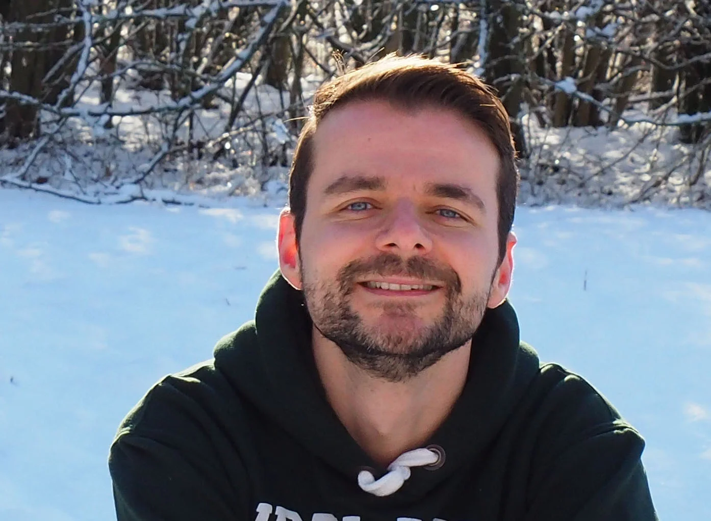
X-ray and neutron imaging applications in soil science Working group member, Internal LINXS Fellow
Dimitrios Floudas, Researcher in Dept. of Biology, Lund University. His research interests are centered on the mechanisms fungi use to transform and decompose organic matter and the evolutionary processes that have shaped those mechanisms. The use of advanced techniques could shed light in processes, such as organic matter decomposition, that play tremendous role in global carbon cycling.
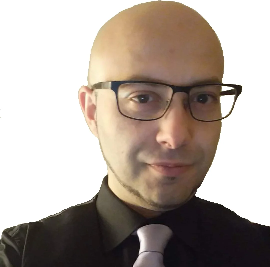
X-ray and neutron imaging applications in soil science Working group member, Internal LINXS Fellow
Luigi Gentile is a researcher at the Biology Department (The MEMEG Unit) of Lund University. His research interests are focused on characterization of soil organic matter and its fungal degradation by means of scattering and spectroscopic techniques. He is experienced in small angle light, X-ray and Neutron scattering.
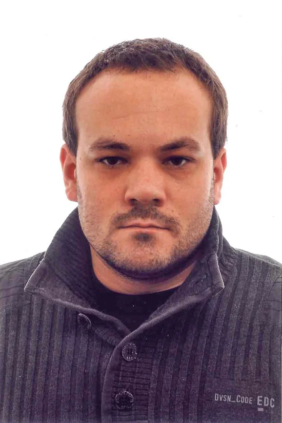
X-ray and neutron imaging applications in soil science Working group member, Internal LINXS Fellow
Michiel Op De Beeck, Researcher at the Dept. of Biology, Lund University. Michiel is a microbiologist with an interest in soil microbial ecology. He employs both infrared microspectroscopy and synchrotron-based X-ray microscopic imaging techniques to study details of the interactions between soil microbes and their environment in situ and at a single cell level.
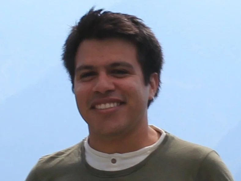
X-ray and neutron imaging applications in soil science Working group member, LINXS Fellow
Luis is a research assistant and Ph.D. candidate at the Chair of Soil Science (TUM-Germany). Luis’s main research interest is the fate and interrelations between carbon and nutrients or contaminants in Tropical and temperate soils. For this Luis applies both sub-micron scale (STXM, µ-FTIR) and bulk (XANES/XRD) synchrotron-based techniques.
imaging wg 4
working group on TBS: tomography of biological samples
Passing from morphology to function is one of the most important challenges that the biomedical community nowadays confronts. Scientists working with biological tissue are most often using visible light microscopy to inspect the samples. The passage from the classical visible light microscopy to tomographic methods should not be described as a simple change of tools. The rapid evolution and the complexity of tomographic methods, deriving from the extensive use of numerical methods and the diversity of beam lights, demands a tight collaboration between biomedical scientists, image analysts, physicists and engineers.
Biological tissues can be studied in two different conditions: ex-vivo and in-vivo, both posing peculiar issues that need to be addressed. In both cases, preservation of their structure during imaging is mandatory. Nonetheless, this is not only a problem of simple preservation of the material. In fact, this already poses an interdisciplinary problem: the interaction between the biological tissue and the type/duration of incident beam. Moreover, the evaluation of tomographic scans requires often competence in mathematical image analysis that exceed the competences of biomedical scientists. In-vivo samples pose an even higher level of challenges. They regard A) the motion of the living parts (hence the necessity of developing solutions for tracking and registering parts of the images), B) the building of different instrument set-ups with life-support for living samples, ranging from simple cell solutions to an entire animal under general anaesthesia.
IPDD WG 3 member and Opportunities in Imaging Working Group member, LINXS Fellow
Martin Bech is Senior Lecturer at the division of Medical Radiation Physics, Clinical Sciences, Lund University. He is interested in X-ray phase contrast and dark-field imaging using grating interferometer, Soft tissue biomedical imaging, Micro-CT imaging using synchrotron radiation and x-ray tubes.
Imaging Working group member, LINXS Fellow
Gaetano Perchiazzi is the director of Respiratory Research at the Hedenstierna Laboratory of Uppsala University, is a medical doctor, specialist in Anesthesia and Intensive Care Medicine; has a PhD in Clinical Physiology on the use of artificial intelligence for extracting information from lung mechanics. Has developed methods for the topographic description of lung elasticity, strain and mechanical energy using computed tomography in animals and humans. He has been principal investigator in different scientific expeditions at the European Synchrotron Radiation Facility (Grenoble, France) for experiments aiming at imaging the micromechanics of lung alveoli in vivo, in conditions simulating clinical mechanical ventilation.
LINXS Co-Director and IPDD WG 3 member
oxana.klementieva@linxs.lu.se
LINXS Co-Director responsible for the focus area of Life Science. Associate Professor, Docent, Co-coordinator Nanobiology & Neuronanoscience at NanoLund and Head of Medical Mirospectrocopy group at the Medical Faculty of Lund University. Research interests include structural changes of amyloid proteins for understanding the nature of neurodegenerative disorders.
IPDD WG 3 member and Imaging Working group member, LINXS Fellow
Hanna Isaksson is a professor in Biomedical Engineering, Lund University. Hanna's reserach area is primarily bone biomechanics and mechanobiology, focusing on functional imaging and material characterization musculoskeletal tissues, primarily bone and tendons. She studies damage and fracture mechanisms and uses a variety of synchrotronbased techniques such as X-ray tomography, scattering and spectrocopy, to understand the link between mechanical performance, structure and composition, on lengthscales from the whole organ down to the building blocks of the tissues.
IPDD WG 3 member and Imaging Working group member, LINXS Fellow
Anja Schmidt-Christensen is a researcher at the Department of Experimental Medical Science at Lund University and the Lund Diabetes Research Center (LUDC). Her research profile and expertise is in the field of immunology of chronic inflammation related to Type-1 Diabetes and fibrosis, with a special focus on advanced imaging methods and working with in vivo models.
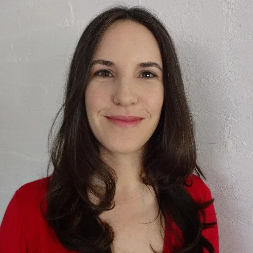
Imaging Working group member, LINXS Fellow
Sonja Aits, Research Team Manager and Associate Senior Lecturer, Faculty of medicine, Lund University, Sweden
imaging wg 5
working group on food science and technology
The full understanding of the complexity of food and food processes requires a combination of techniques and a new toolbox with unsurpassed spatial, chemical and time resolution. MAX IV and ESS will provide unprecedented opportunities for materials sciences and a unique opportunity, both to directly deliver new knowledge required for the understanding of food materials and food related processes but also to provide renewal and thus to strengthen overall science driven development of this major industry. There is an opportunity now to bring leading investigative tools and to attract scientific talent so that major challenges in the food industry and in food science and technology are addressed so as to ensure that Sweden is world-leading in a future industry sector that is technology driven.
However, while the facilities offer advanced technologies for the study of matter it is necessary to build up competences for the use of these techniques in the Swedish food science and food industry sector. It is also of importance to start developing the necessary experimental environments and shape the facilities so that they are suitable to tackle the food specific challenges and development.
Northern Lights on Food website.
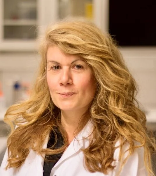
Former Food Core Group Leader & Imaging Working Group Member, LINXS Fellow
Selma Maric, together with Tommy Nylander, launched the Northern Lights on Food initiative, which is now on the way to establish itself into an international network to increase the use of large-scale facilities in food science and food processing. As the Industrial relations Officer at MAX IV Selma is responsible for the areas of life science and soft matter which include food science. Her research has focused on the development of methods for the use of X-ray and neutron-based techniques within life science which has included studies of various lipid-protein assemblies related to both infectious and lifestyle and food-related diseases such as e.g. atherosclerosis. SM will coordinate WG5. Food and health.
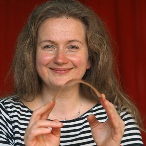
Imaging Working group member, LINXS Fellow
Ramune Kuktaite is an associate professor at the Department for Plant Breeding at the Swedish University of Agricultural Sciences in Alnarp where she focuses on the development of various bioplastics. She is also involved in work focusing on production of protein-rich foods, which can be used as healthy alternatives to meat. She is an avid user of X-ray techniques and represents SLU in the MAX IV University Reference group (URG). Ramune will function as a link to the SLU scientific community to increase both awareness and work on SLUs internal competence building in relation to the facilities.
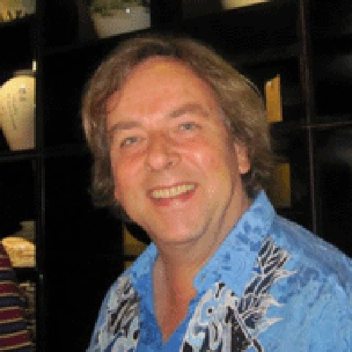
Food Core Group Leader & Dynamics Working Group Dynamics of Membranes and their Constituents Member, Internal LINXS Fellow
Tommy is a professor at the Department of Physical Chemistry at Lund University and an ambassador of the use of X-rays and neutrons in food research. His large network in food science together with his experience in x-ray and neutron scattering on various food related systems is crucial in bringing in the relevant national and international actors to the theme and to the strengthening of food sector using the large-scale infrastructure overall. He is also a member of the management group of NanoLund. Tommy will coordinate WG 4. Food interactions on surfaces.
Food Core Group member & Imaging Working group member, LINXS Fellow
Niklas is a researcher at the Product Design and Perception section at RISE Agrifood and Bioscience. He is also adjunct professor at the department of physics at Chalmers University of Technology. In addition, Niklas is vice director for the VINN Excellence centre SuMo Biomaterials and coordinator for the Soft Microscopy Centre, both close collaborations between RISE and Chalmers. His research generally focuses on microstructure-property relationships in soft heterogeneous bio-based materials such as foods. His network and scientific expertise in microscopy will be crucial for the theme to both develop the working groups and defining the research programs. Together with Stephen Hall, Niklas will coordinate WG3. Structure of food during processing.
Food Core Group member & Imaging Working group member, LINXS Fellow
Anna is a professor at the Division of Pharmaceutical Technology at Chalmers University of Technology, and was a member of the management group of SuMo Biomaterials. Her various research interests include mass transport in soft biomaterials, mechanical properties of biomaterials, and structure and function correlations of polysaccharides. Between 2017 and 2019, she was the Director of Food Science Sweden, a national platform aimed at strengthening and visualising Swedish food science research, and she is expected to be key in raising awareness and building up competence amongst the wider Swedish food science community.
Food Core Group member & Imaging Working Group Member, LINXS Fellow
Lars Nilsson is a professor at Department of Food Technology, Engineering and Nutrition. His research is focused on asymmetrical flow field-flow fractionation (AF4) for the separation of biomolecules and other species of relevance for food systems. Here is one of the leading researchers in the field. He has also large interest in food colloids and the interfacial behavior of food components. He is the main organizer of Food Colloids 2022 in Lund.
imaging wg 6
Working group on quantim: image quantification
The Working Group will enhance the exploitation of complex (mainly 3D) datasets to enable better or new science by giving scientific interpretation to these data. We will create a network consisting of scientists in diverse fields (i.e. biology, geology, material science, palaeontology) and specialists in image analysis.
Quantim has 3 specific aims:
- Establish collaboration (first contact) between data owners and
data scientists
- Train data owners to use analysis tools so they can interpret the
data
- Build a well functioning collection of image analysis tools at
LUNARC
Former LINXS Co-Director and Food WG 3 member, LINXS fellow
Associate professor at the Dept. of Solid Mechanics at the Faculty of Engineering (LTH), where he is also in charge of the 4D-Imaging Lab x-ray tomography facility. Came to Sweden in 2011 after moving from Laboratoire 3R in Grenoble, France.
IPDD WG 3 member and Imaging Core Group Member, LINXS Fellow
Rajmund is a senior scientist, professor at the MAX IV Laboratory (Lund University) and an honorary professor at the Technical University of Denmark. He is a specialist in X-ray physics with emphasis on the enhancement of X-ray imaging for biology and material science applications: development and applications of coherent imaging with improved temporal and spatial resolution.
Imaging Working group member, LINXS Fellow
Vedrana Andersen Dahl is an associate professor at the Department of Applied Mathematics and Computer Science, Technical University of Denmark. She holds degrees in mathematics, multimedia technology, and a Ph.D. degree in geometry processing. Her research interests revolve around geometric models for analysis of volumetric data. This includes volumetric segmentation, tomographic segmentation and methods based on deformable meshes. She developed image analysis tools with application in material science, industrial inspection and biomedicine. Her other research interest involve 2D and 3D texture analysis, analyzing OCT images, and 3D methods for digitizing cultural heritage.
Link to webpage at DTU: http://people.compute.dtu.dk/vand/index.html
Imaging Working group member, LINXS Fellow
Anders Bjorholm Dahl is professor in 3D image analysis at the Department of Applied Mathematics and Computer Science, Technical University of Denmark. He holds a degree in forestry and a Ph.D. degree in computer vision. He is heading the section for Image Analysis and Computer Graphics and QIM: The Center for Quantification of Imaging Data from MAX IV, which has a mission to support quantitative 3D image analysis. His research interests include computer vision and image analysis with a specific focus on learning-based 3D image analysis for structural characterization. This includes volumetric image segmentation, feature- and dictionary-based image representations, graphical models, etc.
Link to webpage at DTU:
https://www.dtu.dk/english/service/phonebook/person?id=32847&tab=2&qt=dtupublicationquery
IMAGING news articles

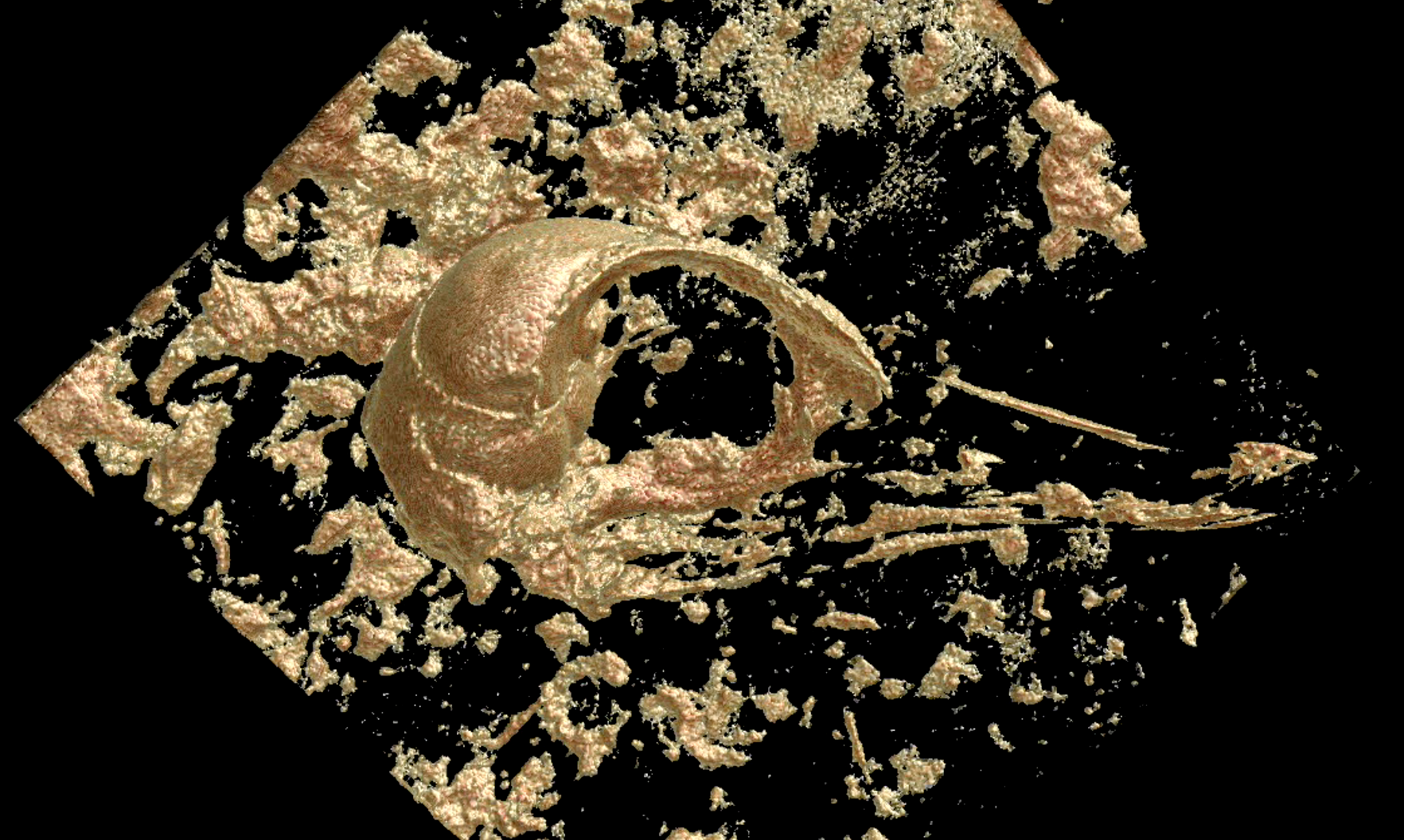
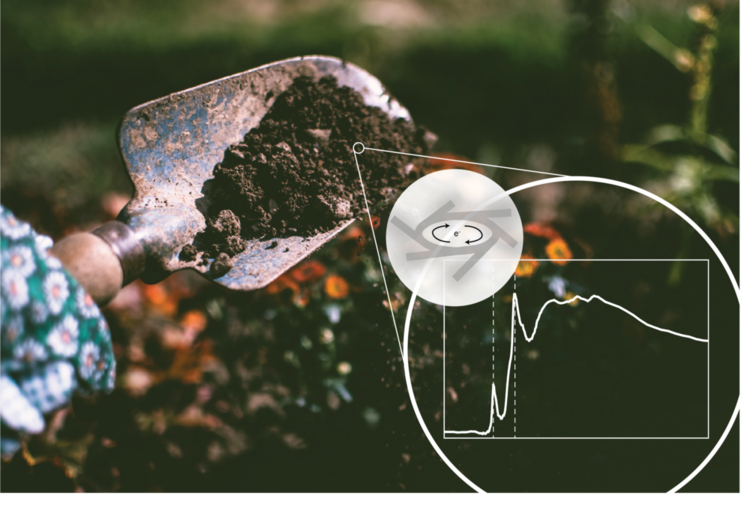
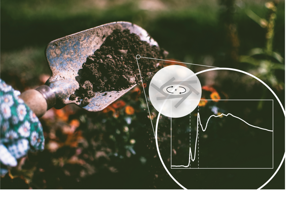
In June, the working group Soil Sciences, is organising its first workshop. In this interview, biologists and working group members, Edith Hammer and Milda Pucetaite talk about what they hope to get out of the event, and the importance of studying soil with the help of new techniques.

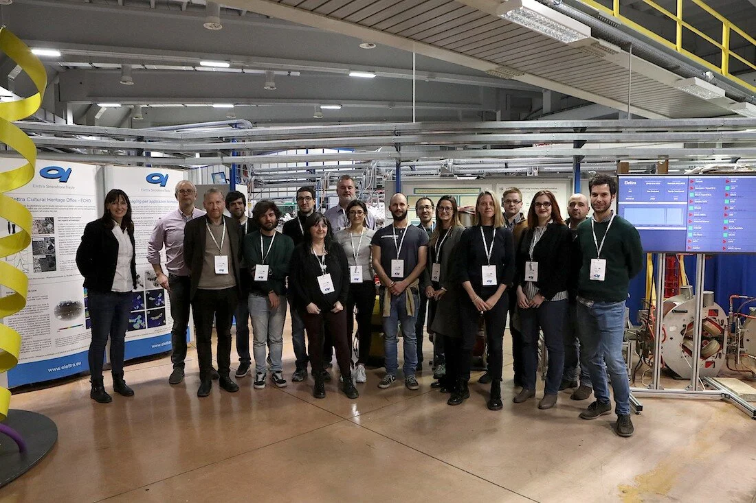
The Quantim workshop in March brought together early researchers and specialists in imaging techniques. The goal was to examine data from five cases ranging from lungs, skin, liver disease and foams, build networks and develop collaborations.
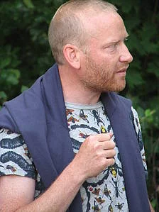
In this interview, archeologists Björn Nilsson, from the Department of Archaeology and Ancient History at Lund University, reflects on his working group’s first symposium and gives some advice to other working groups.
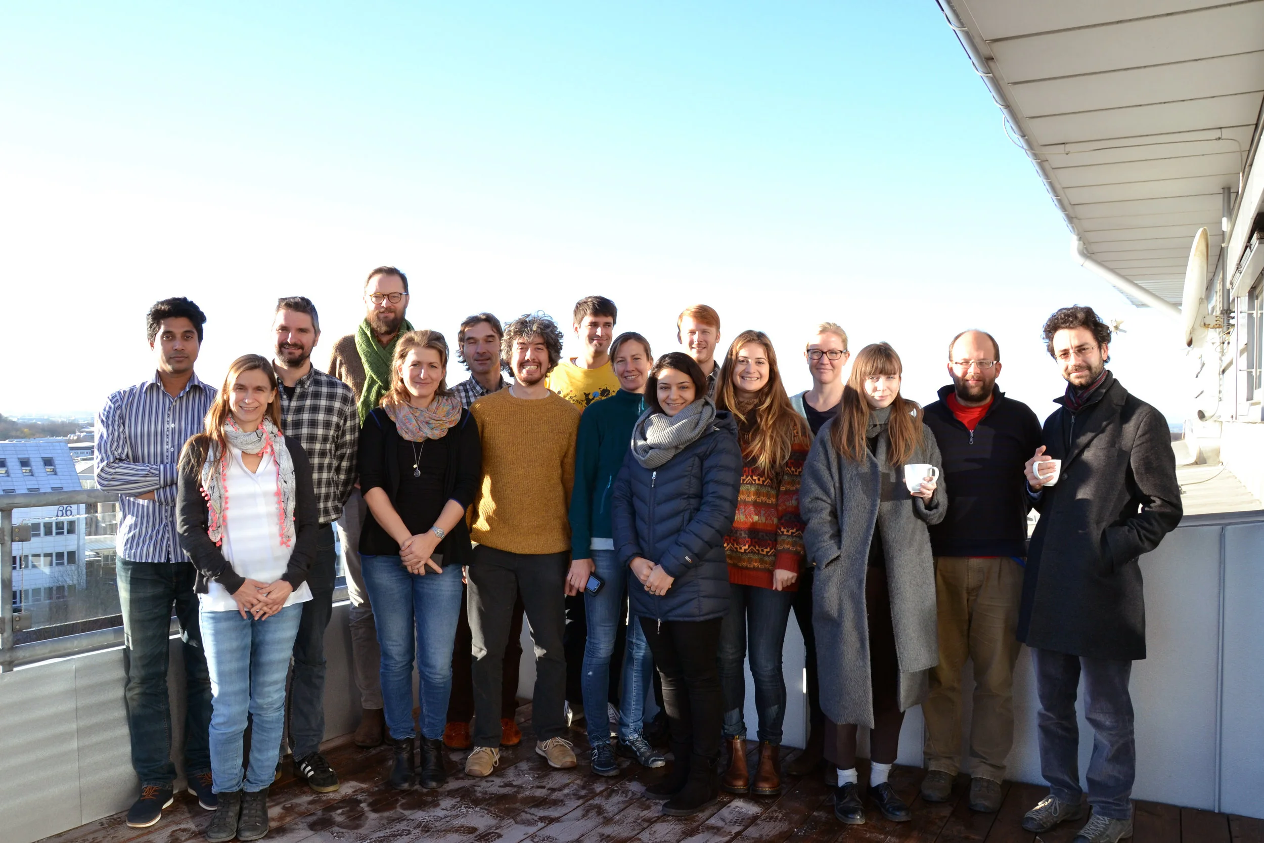
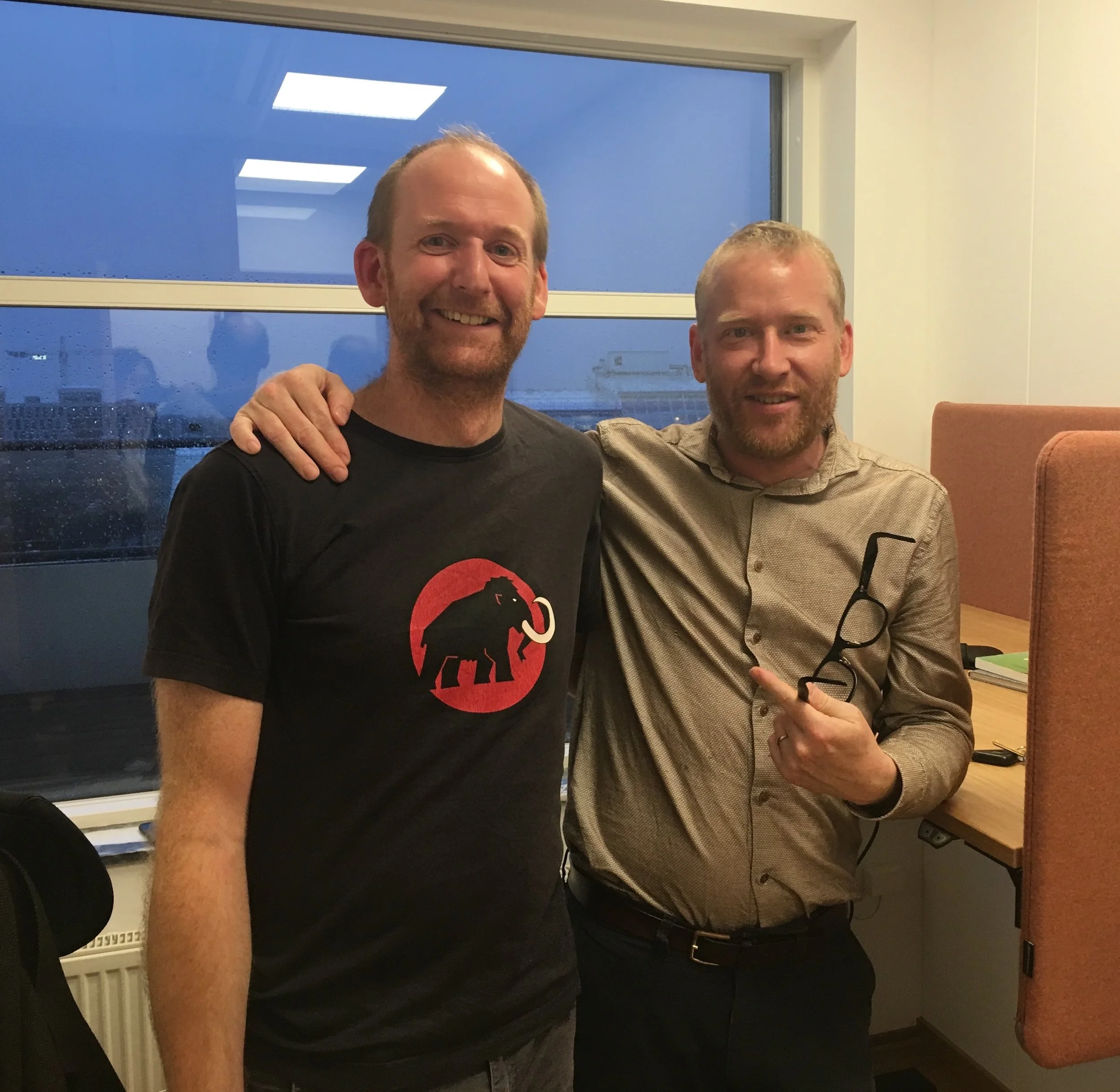

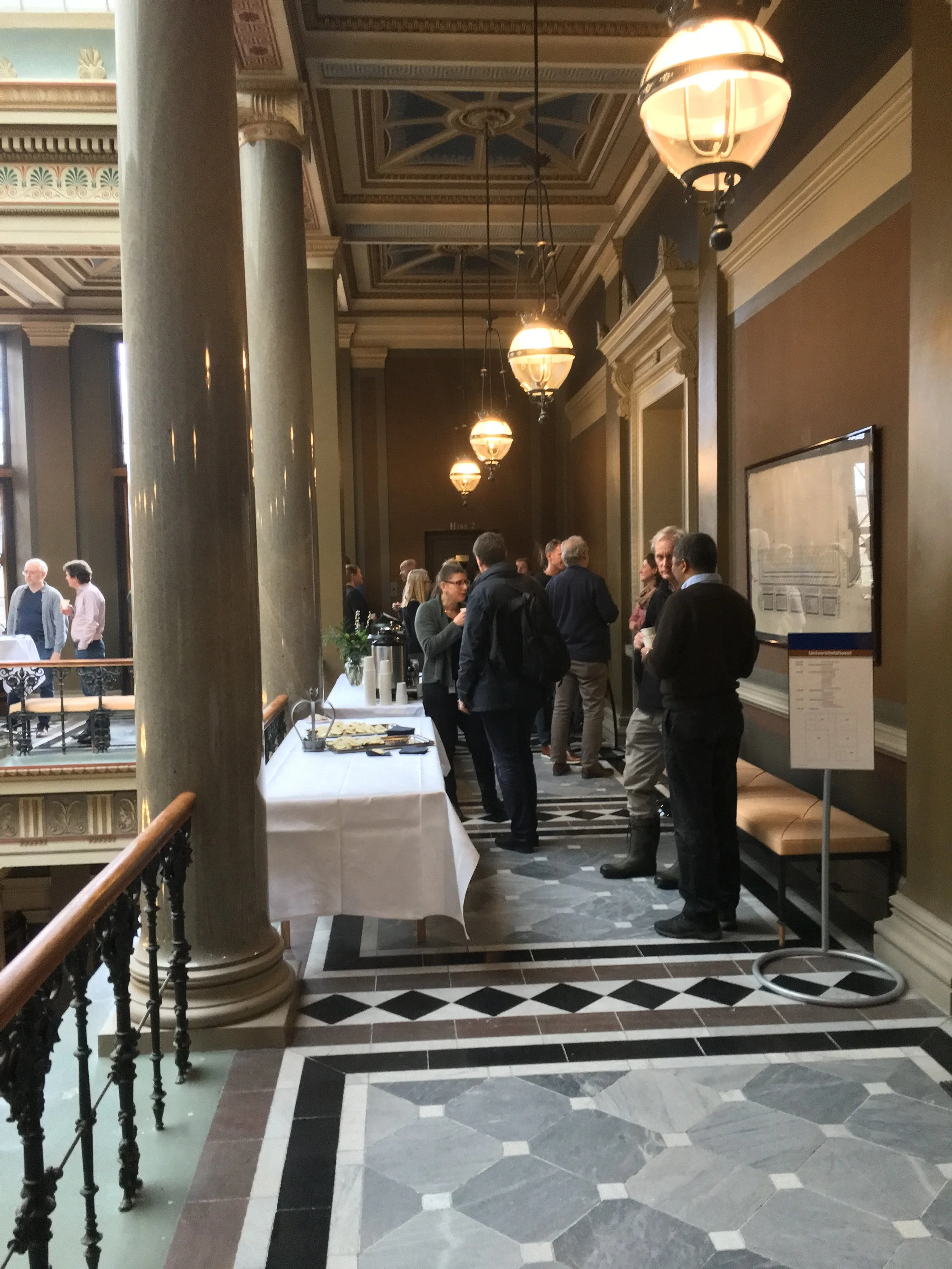
The LINXS Imaging Theme kick-started its activities at the First LINXS-Imaging symposium at the main University building in Lund, 18-19 December 2017.
Imaging Past Events
Welcome to the LINXS Science Day which will focus on the outcomes of the Imaging and Dynamics themes.

Welcome to the 18th Food Colloids Conference: Structure, dynamics and function
10th-13nd of April 2022 in Lund, Sweden
The conference aim is to gather leading international experts from academia and industry to share the latest research and developments in the field of food colloids.
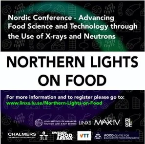
We are proud to announce the second Northern Lights on Food Workshop, June 9-11 2020 in Lund.
Former LINXS Co-Director and Food WG 3 member, LINXS fellow
Associate professor at the Dept. of Solid Mechanics at the Faculty of Engineering (LTH), where he is also in charge of the 4D-Imaging Lab x-ray tomography facility. Came to Sweden in 2011 after moving from Laboratoire 3R in Grenoble, France.