ABOUT
The overarching goal of the Advanced Imaging and Data Analysis (AIDA) theme is the formation of a sustainable, long-term funded Nordic research programme and the establishment of a physical hub and competence platform that can continue developing imaging techniques for the future. In this way, we will establish a network for fostering advanced imaging experts through building excellence and innovation in both research and education.
The Theme builds upon the unique expertise gained in two previous LINXS themes (Imaging and Northern Lights on Food (NLF)) and aims to promote the continued advancement and refinement of both sample preparation and instrumentation, fostering development of scientific and innovation environments to enable cutting-edge deep technology and learning across science areas. This integrated approach of method and science areas will create a long-term benefit to the overall user community both with a continuous stream of new students and new groups, as well as building on the people that have already taken their first steps on the imaging path.
HAPPENING IN THEME
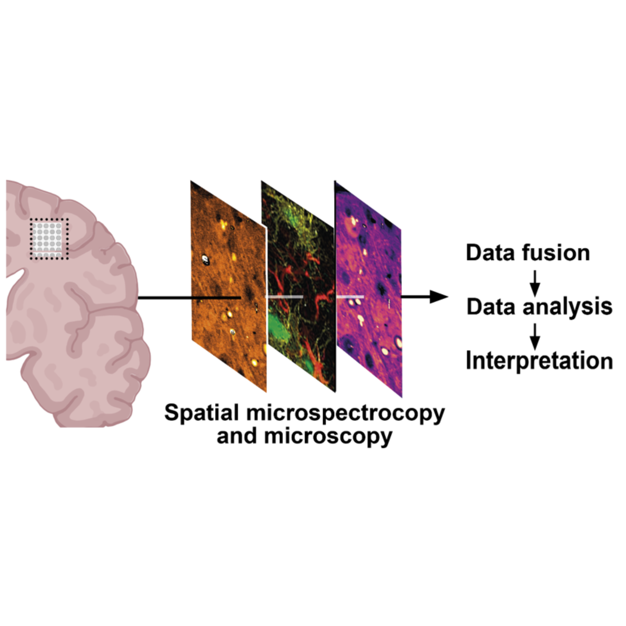
Join us for a two-day workshop on on advanced imaging, spectroscopy, and AI for multiscale brain research, organised by LINXS Themes AIDA and SHINE!
MAX IV and LINXS welcome you to a two-day hackathon on 12-13 Oct 2026 with lectures and hands-on training on PtyPy!
AIDA COMMUNITY LIST SIGNUP
CORE GROUP
Advanced Imaging and Data Analysis: Core Group Co-Leader, Leader of WG 1 – Spectral Nano-imaging, LINXS Fellow
Core responsibility in Theme: Development of Spectral Imaging Pathway
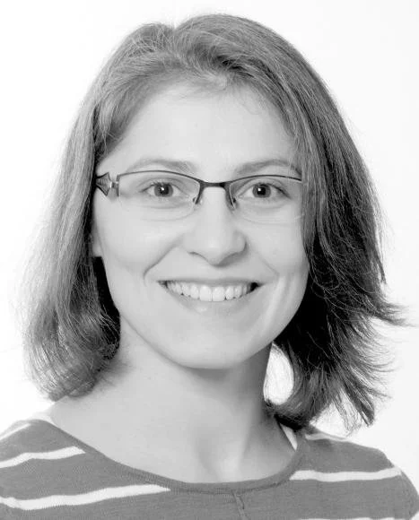
Group Manager, Imaging Beamlines, MAX IV, and Beamline Manager, SoftiMAX, MAX IV, Sweden.
Karina is Group Manager of Imaging Beamlines and beamline scientist at MAX IV with a background in physics and spectroscopy. Her expertise spans a wide range of (synchrotron) techniques and sample systems. With contacts to a wide network of imaging specialists, her aim is to develop the user community, especially towards under-represented science areas. She is also the main organizer of the bi-annual X-ray Microscopy conference, to be held in Lund in August 2024.
Advanced Imaging and Data Analysis: Core Group member, Leader of WG 2 – Phase-contrast micro-tomography, phase retrieval, and reconstruction, LINXS Fellow
Core responsibility in Theme: Data Advancement and Instrument Development
Senior Researcher, Department of Physics, Technical University of Denmark, Denmark, and Senior Scientist, MAX IV and Department of Solid Mechanics, Lund University, Sweden.
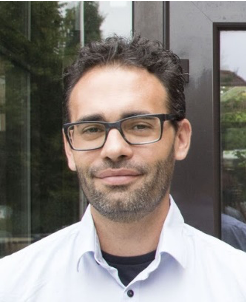
Rajmund is a physicist with experience in building and running synchrotron- and laboratory-based X-ray tomographic instruments. He has an extensive cross disciplinary network with an emphasis on imaging of organic materials.
Advanced Imaging and Data Analysis: Core Group member, Leader of WG 2 – Phase-contrast micro-tomography, phase retrieval, and reconstruction, LINXS Fellow
Core responsibility in Theme: Data Advancement and Instrument Development
Senior lecturer, Division of Solid Mechanics, Faculty of Engineering, Lund University, Sweden.
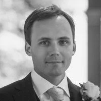
Stephen is a Senior lecturer at the Division of Solid Mechanics at Lund University and former director of LINXS. Stephen’s research covers a wide range of material questions with a focus on understanding the micro scale origins of material mechanics and coupled processes. He works with a wide range of experimental methods, including extensively with x-rays and neutrons and, in particular, imaging and other 2D, 3D and 4D full- field measurement approaches. Stephen runs the 4D Imaging Lab X-ray tomography facility in Lund and heads up the Swedish side of QIM. He has been very much involved in both the ESS and MAX IV projects, including being a member of the imaging and engineering STAP and currently as a SAC member for ESS. He has also been leading the push for a materials imaging beamline at MAX IV.
Advanced Imaging and Data Analysis: Core Group member, Leader of WG 3 – Advanced image processing, image analysis, and visualisation, LINXS Fellow
Core responsibility in Theme: Data Advancement and Instrument Development
Associate professor, Visual Computing, DTU Compute, Technical University of Denmark, Denmark and QIM, MAX IV, Sweden.
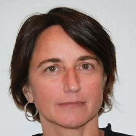
Vedrana is an associate professor at the Department of Applied Mathematics and Computer Science, Technical University of Denmark. She holds degrees in mathematics, multimedia technology, and a Ph.D. degree in geometry processing. Her primary research interest is in using geometric models for the analysis of volumetric data. This includes volumetric segmentation, tomographic segmentation, and methods based on deformable meshes. She developed image analysis tools with applications in material science, industrial inspection, and biomedicine. Her other research interests include quantitative analysis of microstructure, 2D and 3D texture analysis and 3D methods for digitizing cultural heritage.
Advanced Imaging and Data Analysis: Core Group member, Leader of WG 1 – Spectral Nano-imaging, LINXS Fellow
Core responsibility in Theme: Sample preparation and method development
Associate Professor in Molecular Imaging and head of the Medical Microspectroscop Laboratory at Lund University, Sweden.
Oxana is an Associate Professor in Molecular Imaging and head of the Medical Microspectroscopy Laboratory at Lund University. She specialises in molecular imaging. Her research focuses on amyloid proteins associated with neurodegenerative diseases. Oxana achieved significant breakthroughs in infrared multimodal imaging, particularly by pioneering an IR imaging approach for subcellular amyloid detection in AD transgenic neurons (Advanced Science, 2020; JACS 2023). Her group's innovations include the development of correlative nano spectroscopic methods for direct amyloid analysis in neurons, revealing a direct link between β-sheet formation and amyloid-β's pathological effects. From 2021 to 2023, Oxana also served as a co-director of LINXS, where she contributed to the development of the imaging network, with a specific aim of building a multimodal imaging community. In 2023, she furthered her impact by establishing a multiscale IR platform, which features cutting-edge instruments like OPTIR and NanoIR.
Advanced Imaging and Data Analysis: Core Group member, LINXS Fellow
Core responsibility in Theme: Strategy Development, Education and Innovation, Instrument Development
Project Leader, MedMAX, MAX IV, and Development Strategist, Lund University, Sweden.
Selma has a scientific background in the development of scattering methods for studies in health and disease. She has experience from industry, academia, large scale research infrastructure, the Swedish and European funding ecosystem and as the founder of several national and international research networks, educational programs, and innovative start-ups. She founded and developed Northern Lights on Food, a national and international research network, and research theme at the LINXS Institute of Advanced Neutron and X-ray Science. Selma initiated the national collaboration Infralife joining MAX IV, ESS and Scilifelab to open and broaden the use of infrastructure in Life Science to both academia, industry, and health care. Developed PRISMAS, a national PhD program training 40 PhD students joining nine universities and 43 associated partners. She is project leader for the development a life science beamline and corresponding user community at MAX IV. Currently, Selma leads the implementation phase for the establishment of “Swedish Platforms for Advanced Infrastructure for Research, Innovation and Technology (SPIRIT)”, a national technical park function surrounding MAX IV and ESS.
Advanced Imaging and Data Analysis: Core Group member, Leader of ASG in Palaeontology and Geotech, LINXS Fellow
Core responsibility in Theme: Education, competence, and Innovation
Associate Professor, Department of Organismal Biology, Uppsala University, Sweden.
Sophie is an Associate professor at Uppsala University. She has a scientific background in evolutionary biology and palaeontology, and has developed virtual palaeohistology during her postdoc in collaboration with colleagues from the European Synchrotron Radiation Facility (ESRF) for submicron investigations of fossils using X-ray microtomography. She is PI of an ERC Consolidator and several VR grants developing the use of synchrotron imaging for histological investigations of fossils at the fish-to-tetrapod transition. Sophie has been an invited speaker at the World Meeting on Heritage, Sciences and Technologies, in Paris (2019), ESRF-EBS workshop on Cultural and Natural Heritage, in Grenoble (2020) and LINXS Symposium and Hackathon IPDD theme – 3D Histology with Synchrotron Microtomography, in Lund (2023) for promoting synchrotron 3D imaging in cultural fields. Invited to teach in the following PhD summer school/courses São Paulo School of Advanced Science (ESPCA), Synchrotron of Campinas, Brazil (2011), RÅC International Summer School (RACIRI), Stockholm, Sweden (2014), Synchrotron Radiation in Natural Sciences, Uppsala University, Sweden (2016-2018). She is a member of the University Reference Group at MAX IV (Uppsala University scientific advisor in Life Science) and board member of the Center for Photon Science at Uppsala University. She has been engaged in proposal and cross-cut review panels at the ESRF for life science, palaeontology, and cultural heritage.
WORKING GROUPS FOR AIDA
AIDA working group 1
Spectral Nano-imaging
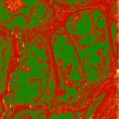
In this working group, the chemical speciation takes centre stage in combination with ultimate resolution methods in chemical imaging. This means, for instance, identifying elements with X-ray Fluorescence, but also disentangling different chemical species of the same element with Scanning Transmission X- ray Microscopy and complementary IR-microscopies. Mostly conducted in 2D, the 3rd dimension is formed by the spectral component. The big advantage is that these techniques require no labelling and instead purely work with the composition or chemistry inherent to the sample, which is therefore relevant for a wide variety of scientific fields. Over the past five years, nano-imaging techniques have come to fruition at MAX IV, with the addition of new operational beamlines and end-stations, and now the time is right for e.g., the development of more streamlined data analysis approaches and assistance with sample preparation for experiments. A separate aim is to reach out to experts in laboratory-based, high-resolution imaging techniques, including optical (fluorescence) methods and spatial proteomics as well as electron-based methods such as TEM and STM while bridging to working group 2. This integration expands the scope of synchrotron experiments, thus providing a comprehensive nano-imaging toolkit.
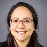
Advanced Imaging and Data Analysis: Core Group Co-Leader, Leader of WG 1 – Spectral Nano-imaging, LINXS Fellow
Core responsibility in Theme: Development of Spectral Imaging Pathway
Group Manager, Imaging Beamlines, MAX IV, and Beamline Manager, SoftiMAX, MAX IV, Sweden.
Karina is Group Manager of Imaging Beamlines and beamline scientist at MAX IV with a background in physics and spectroscopy. Her expertise spans a wide range of (synchrotron) techniques and sample systems. With contacts to a wide network of imaging specialists, her aim is to develop the user community, especially towards under-represented science areas. She is also the main organizer of the bi-annual X-ray Microscopy conference, to be held in Lund in August 2024.
Advanced Imaging and Data Analysis: Core Group member, Leader of WG 1 – Spectral Nano-imaging, LINXS Fellow
Core responsibility in Theme: Sample preparation and method development
Associate Professor in Molecular Imaging and head of the Medical Microspectroscop Laboratory at Lund University, Sweden.
Oxana is an Associate Professor in Molecular Imaging and head of the Medical Microspectroscopy Laboratory at Lund University. She specialises in molecular imaging. Her research focuses on amyloid proteins associated with neurodegenerative diseases. Oxana achieved significant breakthroughs in infrared multimodal imaging, particularly by pioneering an IR imaging approach for subcellular amyloid detection in AD transgenic neurons (Advanced Science, 2020; JACS 2023). Her group's innovations include the development of correlative nano spectroscopic methods for direct amyloid analysis in neurons, revealing a direct link between β-sheet formation and amyloid-β's pathological effects. From 2021 to 2023, Oxana also served as a co-director of LINXS, where she contributed to the development of the imaging network, with a specific aim of building a multimodal imaging community. In 2023, she furthered her impact by establishing a multiscale IR platform, which features cutting-edge instruments like OPTIR and NanoIR.
Advanced Imaging and Data Analysis: member of WG 1 – Spectral Nano-imaging, LINXS Fellow
Beamline scientist, Diamond Light Source, United Kingdom.
Advanced Imaging and Data Analysis: member of WG 1 – Spectral Nano-imaging, LINXS Fellow
Beamline manager, Soleil Synchrotron, France.
Advanced Imaging and Data Analysis: member of WG 1 – Spectral Nano-imaging, LINXS Fellow
Lecturer, Nanoscale Bioscience, Keele University, United Kingdom.
Advanced Imaging and Data Analysis: member of WG 1 – Spectral Nano-imaging, LINXS Fellow
Professor Emeritus, Chemistry & Chemical Biology, Department of Chemistry, McMaster University, Canada.
Advanced Imaging and Data Analysis: member of WG 1 – Spectral Nano-imaging and WG3 – Advanced image processing, image analysis, and visualisation, LINXS Fellow
Associate professor and researcher, Computational Biology and Biological Physics, Centre for Environmental and Climate Science, Faculty of Science, Lund University and Application Expert, CIPA and InfraVis, Sweden.
AIDA WORKING GROUP 2
Micro-tomographic Imaging
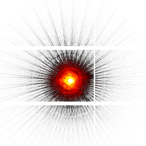
Phase-contrast micro-computed tomography is an imaging modality that has evolved rapidly in the past few decades. The method exploits the wave-like behaviour of x-rays and neutrons, providing images with superior contrast in comparison to standard attenuation-based imaging. Due to the coherent properties of synchrotron radiation, phase-contrast imaging has been implemented at all synchrotron imaging beamlines, but even with neutron imaging it is possible to obtain phase-contrast images by use of optical gratings to produce beam interference. In these grating interferometers, it is even possible to extract so-called dark-field images based on the scattering in the sample. Such grating interferometers are even implemented at laboratory x-ray micro-tomography setups, along with propagation-based phase-contrast imaging.
The method experts in WG2 of the AIDA theme work with micro-tomographic imaging at synchrotrons, at neutron facilities, at free-electron laser facilities, and at laboratory-based x-ray sources both in academia and industry. Networking activities internally in the WG2 will provide cross fertilization for method optimization at different instruments and provide a strong basis of competence for the Advanced Study Groups.
Advanced Imaging and Data Analysis: Core Group member, Leader of WG 2 – Phase-contrast micro-tomography, phase retrieval, and reconstruction, LINXS Fellow
Core responsibility in Theme: Data Advancement and Instrument Development
Senior Researcher, Department of Physics, Technical University of Denmark, Denmark, and Senior Scientist, MAX IV and Department of Solid Mechanics, Lund University, Sweden.
Rajmund is a physicist with experience in building and running synchrotron- and laboratory-based X-ray tomographic instruments. He has an extensive cross disciplinary network with an emphasis on imaging of organic materials.
Advanced Imaging and Data Analysis: Core Group member, Leader of WG 2 – Phase-contrast micro-tomography, phase retrieval, and reconstruction, LINXS Fellow
Core responsibility in Theme: Data Advancement and Instrument Development
Senior lecturer, Division of Solid Mechanics, Faculty of Engineering, Lund University, Sweden.
Stephen is a Senior lecturer at the Division of Solid Mechanics at Lund University and former director of LINXS. Stephen’s research covers a wide range of material questions with a focus on understanding the micro scale origins of material mechanics and coupled processes. He works with a wide range of experimental methods, including extensively with x-rays and neutrons and, in particular, imaging and other 2D, 3D and 4D full- field measurement approaches. Stephen runs the 4D Imaging Lab X-ray tomography facility in Lund and heads up the Swedish side of QIM. He has been very much involved in both the ESS and MAX IV projects, including being a member of the imaging and engineering STAP and currently as a SAC member for ESS. He has also been leading the push for a materials imaging beamline at MAX IV.
Advanced Imaging and Data Analysis: member of WG 2 – Phase-contrast micro-tomography, phase retrieval, and reconstruction and WG3 – Advanced image processing, image analysis, and visualisation, LINXS Fellow
Researcher, Department of Experimental Medical Science, Faculty of Medicine, Lund University; Node Coordinator, InfraVis; Coordinator, CIPA; Associate Researcher, QIM, MAX IV; Sweden.
Advanced Imaging and Data Analysis: member of WG 2 – Phase-contrast micro-tomography, phase retrieval, and reconstruction, LINXS Fellow
Senior Researcher, Department of Applied Mathematics and Computer Science, Technical University of Denmark, Denmark.
Advanced Imaging and Data Analysis: member of WG 2 – Phase-contrast micro-tomography, phase retrieval, and reconstruction, LINXS Fellow
Leader Applied Materials, Swiss Spallation Neutron Source (SINQ), Paul Scherrer Institute (PSI), Switzerland.
Advanced Imaging and Data Analysis: member of WG 2 – Phase-contrast micro-tomography, phase retrieval, and reconstruction, LINXS Fellow
Associate senior lecturer, Synchrotron Radiation Research, Lund University, Sweden.
Advanced Imaging and Data Analysis: member of WG 2 – Phase-contrast micro-tomography, phase retrieval, and reconstruction and WG3 – Advanced image processing, image analysis, and visualisation, LINXS Fellow
Research scientist, Centre National de Recherche Scientifique (CNRS), TIMC Laboratory, France.
Advanced Imaging and Data Analysis: member of WG 2 – Phase-contrast micro-tomography, phase retrieval, and reconstruction, LINXS Fellow
Technology Specialist, Exiscope AB, Sweden.
Advanced Imaging and Data Analysis: member of WG 2 – Phase-contrast micro-tomography, phase retrieval, and reconstruction, LINXS Fellow
Postdoctoral researcher, ForMAX beam line, MAX IV, Sweden.
AIDA WORKING GROUP 3
Advanced image processing
AIDA advanced study GROUP:
tissue and in vivo imaging
AIDA advanced study GROUP:
food and biotech
AIDA advanced study GROUP:
paleontology and geotech
This working group is a fundamental layer between the acqiusition of raw data and scientific interpretation, dealing with specific data-oriented goals while facing a cross-disciplinary challenge. Besides focused activities inside the working group to advance the use of e.g. machine learning, VR and AR for imaging methods, cross-working group initiatives, such as hackathons and visualization-themed workshops are also foreseen, organized around the central themes provided by the Advanced Study Groups.
The primary emphasis in this working group will be on innovative approaches for extracting multiscale information from X-ray/neutron imaging data, visualizing results in an comprehensive way and delivering this whole process as a user-friendly environment. Additionally, we aim to foster collaboration among Imaging method users to share their expertise to educate and mentor the next generation of scientists.
Advanced Imaging and Data Analysis: Core Group member, Leader of WG 3 – Advanced image processing, image analysis, and visualisation, LINXS Fellow
Core responsibility in Theme: Data Advancement and Instrument Development
Associate professor, Visual Computing, DTU Compute, Technical University of Denmark, Denmark and QIM, MAX IV, Sweden.
Vedrana is an associate professor at the Department of Applied Mathematics and Computer Science, Technical University of Denmark. She holds degrees in mathematics, multimedia technology, and a Ph.D. degree in geometry processing. Her primary research interest is in using geometric models for the analysis of volumetric data. This includes volumetric segmentation, tomographic segmentation, and methods based on deformable meshes. She developed image analysis tools with applications in material science, industrial inspection, and biomedicine. Her other research interests include quantitative analysis of microstructure, 2D and 3D texture analysis and 3D methods for digitizing cultural heritage.
Advanced Imaging and Data Analysis: member of WG 1 – Spectral Nano-imaging and WG3 – Advanced image processing, image analysis, and visualisation, LINXS Fellow
Associate professor and researcher, Computational Biology and Biological Physics, Centre for Environmental and Climate Science, Faculty of Science, Lund University and Application Expert, CIPA and InfraVis, Sweden.
Advanced Imaging and Data Analysis: member of WG 2 – Phase-contrast micro-tomography, phase retrieval, and reconstruction and WG3 – Advanced image processing, image analysis, and visualisation, LINXS Fellow
Researcher, Department of Experimental Medical Science, Faculty of Medicine, Lund University; Node Coordinator, InfraVis; Coordinator, CIPA; Associate Researcher, QIM, MAX IV; Sweden.
Advanced Imaging and Data Analysis: member of WG 2 – Phase-contrast micro-tomography, phase retrieval, and reconstruction and WG3 – Advanced image processing, image analysis, and visualisation, LINXS Fellow
Research scientist, Centre National de Recherche Scientifique (CNRS), TIMC Laboratory, France.
Advanced Imaging and Data Analysis: member of WG3 – Advanced image processing, image analysis, and visualisation, LINXS Fellow
Application Specialist, Exiscope AB, Sweden.
Advanced Imaging and Data Analysis: member of WG3 – Advanced image processing, image analysis, and visualisation, LINXS Fellow
Head of QIM, MAX IV, Sweden, and Professor, 3D image analysis and Head of Section for Visual Computing, DTU Compute, Technical University of Denmark, Denmark.
Advanced Imaging and Data Analysis: member of WG3 – Advanced image processing, image analysis, and visualisation, LINXS Fellow
Professor, Image Analysis, Computational Modelling and Geometry, Department of Computer Science, University of Copenhagen, Denmark.
Tissues are inherently three dimensional, yet the traditional way of analyzing different types of biological and pathological processes in tissue is to section it and then apply various methods like histology, immunohistochemistry and in situ hybridization on the thin 2D slices. However, many organs contain structures which may be difficult to understand from a 2D slice. This is especially the case when the tissue organization has been disrupted by disease processes. During recent years, life science researchers from different fields have started to explore the use of synchrotron-based X-ray imaging methods such as phase-contrast micro-CT, X-ray fluorescence mapping and scanning SAXS/WAXS for tissue characterization. Since the imaging can be performed in a non-invasive way, valuable tissue from biobanks can be scanned and analyzed. Currently there is a great need for knowledge exchange between researchers from different fields to make sure that novel techniques are shared and used in the most efficient way.
Advanced Imaging and Data Analysis: Leader of ASG on tissue and in vivo imaging
Physician Scientist, Skåne University Hospital and Lund University, Sweden.
Advanced Imaging and Data Analysis: Leader of ASG on tissue and in vivo imaging
Professor, Department of Chemistry, Aarhus University, Denmark.
Biomass and derived food materials are complex assemblies of biopolymers and small molecules, in terms of their molecular composition and multiscale organization from the molecular to the macroscopic level. Moreover, all processing steps in industrial biotechnology and food (bio)technology influence this multiscale structure, and they often require time resolved analyses to follow their development. Considering the food sector, all steps in the preparation of the food products, from the raw materials, extraction of suitable ingredients, processing and storage, affect the sensory properties of the food we ingest as consumers, the bioavailability of the nutrients through the gastrointestinal tract, and the derived health benefits. Therefore, the integration of imaging techniques coupled with X-ray and neutron techniques is required to understand the composition and structural properties of food materials in all steps of their cycle. Efforts should be made to develop new tools for image processing and analysis of the vast amount of data generated by such experiments with the help of deep machine learning and artificial intelligence tools.
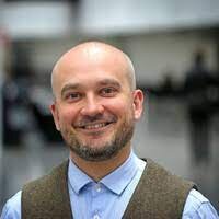
Advanced Imaging and Data Analysis: Leader of ASG in food and biotech
Professor and Head of Department, Division of Glycoscience, KTH Royal Institute of Technology, Sweden.
Advanced Imaging and Data Analysis: Leader of ASG in food and biotech
Professor, Department of Food Science, Aarhus University, Denmark.
Geological, fossil and archaeological samples form a large component of our cultural heritage and, in that sense, they can be far too rare and precious to be sectioned or sampled. Three-dimensional imaging has been developed to overcome this limitation and allow for microstructural and chemical analysis of this crucial material. However, this is not without challenges. These samples are often dense and comprise embedded metallic oxides that strongly react with X-ray and create artifacts. We have formed a community of synchrotron scientists and synchrotron users (with key members in the Nordic countries) to develop and apply synchrotron X-ray imaging on these samples. We propose to introduce the methods of X-ray (micro-)tomography, diffraction tomography and fluorescence to the communities of Geotech and Cultural Heritage to increase the visibility of these techniques to the study of our Nordic history.

Advanced Imaging and Data Analysis: Core Group member, Leader of ASG in Palaeontology and Geotech, LINXS Fellow
Core responsibility in Theme: Education, competence, and Innovation
Associate Professor, Department of Organismal Biology, Uppsala University, Sweden.
Sophie is an Associate professor at Uppsala University. She has a scientific background in evolutionary biology and palaeontology, and has developed virtual palaeohistology during her postdoc in collaboration with colleagues from the European Synchrotron Radiation Facility (ESRF) for submicron investigations of fossils using X-ray microtomography. She is PI of an ERC Consolidator and several VR grants developing the use of synchrotron imaging for histological investigations of fossils at the fish-to-tetrapod transition. Sophie has been an invited speaker at the World Meeting on Heritage, Sciences and Technologies, in Paris (2019), ESRF-EBS workshop on Cultural and Natural Heritage, in Grenoble (2020) and LINXS Symposium and Hackathon IPDD theme – 3D Histology with Synchrotron Microtomography, in Lund (2023) for promoting synchrotron 3D imaging in cultural fields. Invited to teach in the following PhD summer school/courses São Paulo School of Advanced Science (ESPCA), Synchrotron of Campinas, Brazil (2011), RÅC International Summer School (RACIRI), Stockholm, Sweden (2014), Synchrotron Radiation in Natural Sciences, Uppsala University, Sweden (2016-2018). She is a member of the University Reference Group at MAX IV (Uppsala University scientific advisor in Life Science) and board member of the Center for Photon Science at Uppsala University. She has been engaged in proposal and cross-cut review panels at the ESRF for life science, palaeontology, and cultural heritage.
Advanced Imaging and Data Analysis: member of ASG in Palaeontology and Geotech
Chair, Experimental Mechanics, Luleå University of Technology, Sweden.
AIDA news articles
AIDA upcoming events
Join us for a two-day workshop on on advanced imaging, spectroscopy, and AI for multiscale brain research, organised by LINXS Themes AIDA and SHINE!
MAX IV and LINXS welcome you to a two-day hackathon on 12-13 Oct 2026 with lectures and hands-on training on PtyPy!
AIDA past events
Join us for a mini hackathon on 3D data segmentation, featuring a demonstration of the QIM3d tool by William Laprade from DTU Compute, followed by a hands-on session using your own synchrotron or lab-based X-ray tomography data.
Hands-on training and advanced applications of Optical Photothermal Infrared (OPTIR) imaging for structural biology and protein aggregation research.
Advanced Imaging and Data Analysis: Core Group Leader, LINXS Fellow
Core responsibility in Theme: Education, Competence, and Innovation
Associate Professor, Department of Medical Radiation Physics, Lund University, Sweden.
Martin is Associate Professor at Medical Radiation Physics at Lund University. He has a scientific background in X-ray physics and specialized in X-ray imaging method development. Martin has a strong track record of grating-based X-ray interferometry for X-ray phase-contrast and X-ray dark-field imaging for imaging of organic materials at synchrotron radiation facilities, at conventional X-ray sources and at neutrons facilities. He has been an active member of the LINXS community since the start of the Imaging theme and is playing an active role in the development of a new beamline at MAX IV dedicated to tomography and imaging of organic materials. He has more than 10 years of experience in undergraduate teaching of ionising radiation and was co-founder and leader of the graduate research school “Imaging of 3D structures” organised across the faculties of science, engineering, and medicine at Lund University 2014-2019. In addition to user experience at multiple European synchrotrons, Martin is engaged in the proposal review panels at PETRA III and MAX IV. He has received research funding from the Swedish Foundation for Strategic Research, from the Swedish Research council, and is currently PI on an ERC consolidator grant developing X-ray imaging instrumentation for bio-medical research.